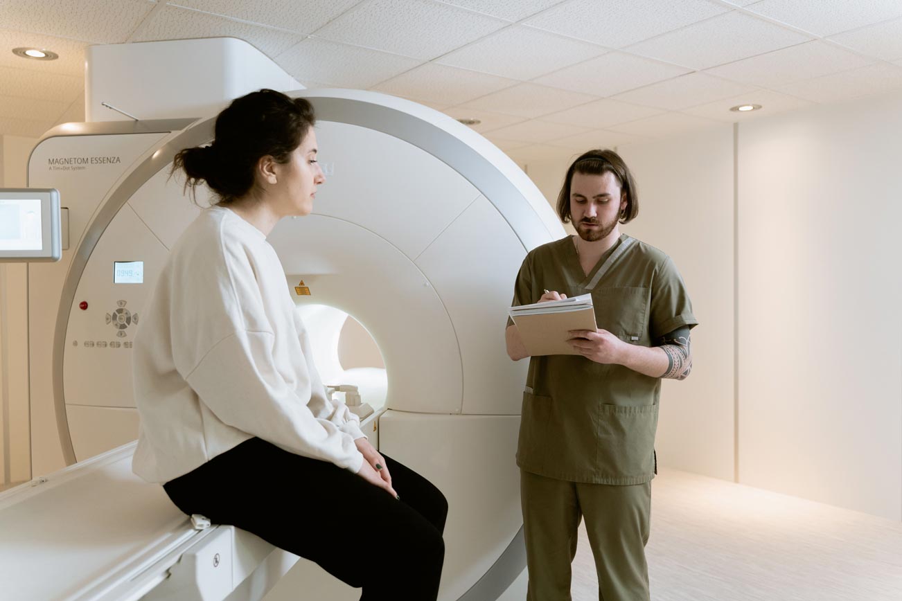
Pregnancy often changes the way we think about medical investigations. Something that might previously have felt routine, such as an X-ray or CT scan, can suddenly raise an entirely different set of questions when there is a baby developing inside you.
Is the radiation exposure dangerous? Could it affect your baby’s development? Should you refuse an X-ray if you are pregnant? Why would a doctor recommend a CT scan when an ultrasound is available?
These are reasonable questions, particularly because medical imaging is often discussed in terms of radiation exposure without explaining how that exposure is measured, managed or minimised.
The good news is that modern medical imaging during pregnancy is guided by carefully established safety principles.
One of the most important is ALARA – As Low As Reasonably Achievable.
ALARA is a radiation protection principle that guides healthcare professionals in minimising unnecessary radiation exposure while still obtaining the diagnostic information needed to make appropriate medical decisions.
During pregnancy, this means carefully considering which imaging technique is most appropriate, whether the investigation is necessary, how much radiation may reach the developing baby and what adjustments can be made to reduce exposure without compromising the quality of the examination.
Importantly, protecting your baby does not always mean avoiding medical imaging. Sometimes, identifying a potentially serious condition early is the safest decision for both mother and baby.
What Does ALARA Actually Mean?
ALARA, as mentioned, stands for As Low As Reasonably Achievable.
It is an internationally recognised radiation protection principle used in medical imaging, radiology, nuclear medicine and other environments where ionising radiation is involved.
The principle is straightforward:
Radiation exposure should be kept as low as reasonably possible while still achieving the medical purpose of the investigation.
This does not mean that doctors simply use the lowest possible radiation setting for every scan. An image that is too poor to interpret may need to be repeated, potentially exposing the patient to more radiation than a properly planned examination would have required.
Instead, ALARA involves finding the right balance between:
- Obtaining sufficient diagnostic information.
- Avoiding unnecessary or repeated examinations.
- Using the lowest appropriate radiation dose.
- Protecting surrounding tissues and organs.
- Considering the patient’s individual circumstances, including pregnancy.
During pregnancy, this requires particular attention because the developing embryo or fetus may be sensitive to certain effects of radiation, depending on the dose received and the stage of its development.
The ALARA principle is built around three important considerations:
Justification
Is the investigation medically necessary, and will the results influence clinical management?
Optimisation
Can the investigation be performed using a technique or protocol that reduces radiation exposure without compromising diagnostic accuracy?
Dose limitation
Are unnecessary exposures being avoided, and is the radiation dose being kept within appropriate safety parameters?
These considerations help doctors make informed decisions on whether ionising or non-ionising imaging scans are the better course of action rather than relying on a blanket rule that all radiation is dangerous during pregnancy.
Understanding the Difference Between Ionising and Non-Ionising Imaging
Not all medical imaging uses radiation in the same way.
One of the most important distinctions when discussing pregnancy imaging is the difference between ionising and non-ionising radiation.
Ionising radiation carries enough energy to remove electrons from atoms. This can potentially damage biological tissue and DNA. The extent of any biological effect depends on factors such as the radiation dose, the tissue exposed and the duration or pattern of exposure.
Non-ionising imaging techniques use different physical processes and do not produce the same type of ionisation.
Understanding this distinction makes it easier to understand why certain investigations are preferred during pregnancy.
Medical Imaging Techniques Using Ionising Radiation
There are three very common medical imaging techniques used in medicine that use ionising radiation.
They are as follows:
X-rays
X-rays use ionising radiation to create images of structures inside the body, particularly bones, the chest and certain abdominal structures.
They are commonly used to investigate fractures, chest infections and other medical conditions.
The amount of radiation reaching the baby depends largely on whether the uterus is within or close to the primary X-ray radiation beam. For example, a chest X-ray exposes the fetus to extremely low levels of scattered radiation because the uterus is outside the area being directly imaged. Whereas, an X-ray of the pelvis procures more risk to the fetus because of its direct proximity to the area.
The decision to get an x-ray done will be determined by the doctor primarily based on the severity of the need and requirements of the patient’s situation.
Computed Tomography (CT) Scans

CT examinations can involve more radiation than conventional X-rays, therefore, the scan area and imaging protocol require careful consideration during pregnancy.
CT scans use multiple X-ray measurements to produce detailed cross-sectional images of the body. Therefore, they are particularly useful when doctors need to investigate internal bleeding, serious injuries, blood clots, appendicitis or other potentially urgent conditions.
The doctor in charge will determine the need for one. A necessary CT scan is generally not avoided simply because a patient is pregnant. However, it is important to remember that different situations require different levels of care.
Fluoroscopy and Nuclear Medicine

Fluoroscopy uses continuous or pulsed X-ray imaging to produce moving images of internal structures. It is sometimes used during specialised procedures, including certain vascular and gastrointestinal investigations.
Nuclear medicine uses small amounts of radioactive substances, known as radiopharmaceuticals, to assess organ function and identify particular diseases.
These techniques require additional consideration during pregnancy because radiation exposure may involve more than a single image or may include radioactive substances that circulate within the body.
Where possible, alternatives are considered, particularly when the investigation can safely wait until after delivery.
Imaging Techniques That Do Not Use Ionising Radiation
The distinction to ionising imaging techniques is important.
Non-ionising does not automatically mean that an investigation should be performed without clinical reason. Every imaging examination should have a clear medical purpose.
There are two very common medical imaging techniques used in medicine that do not use ionising radiation.
They are as follows:
Ultrasound

Ultrasound uses high-frequency sound waves to create images of the uterus, placenta and developing baby.
It does not use ionising radiation and is the primary imaging technique used throughout routine antenatal care.
It is also useful for investigating abdominal pain, bleeding, pregnancy location and several other obstetric concerns. It is the more common choice, as opposed to the CT scan.
Magnetic Resonance Imaging (MRI)

MRI uses strong magnetic fields and radiofrequency energy to produce detailed images of internal organs and soft tissues.
It does not use ionising radiation and may be particularly useful when ultrasound cannot provide sufficient information.
MRI can be more commonly considered during pregnancy when clinically indicated, although certain precautions apply, particularly regarding contrast agents and how they may affect the patient, the baby and the pregnancy going forward.
How Does Radiation Actually Affect a Developing Baby?
The developing baby is more sensitive to certain effects of ionising radiation than an adult because cells are dividing and developing rapidly.
During pregnancy, these cells are responsible for forming the baby’s organs, tissues and major body systems. Exposure to sufficiently high levels of radiation can interfere with normal cell development, potentially affecting how certain structures form and function.
However, the presence of radiation exposure does not automatically mean that harm has occurred. The potential effects depend on several factors, particularly the amount of radiation absorbed by the developing baby, the area of the mother’s body being examined and the stage of pregnancy at the time of exposure.
The possibility of harm depends on several factors, including:
- The total radiation dose received.
- The area of the mother’s body being examined.
- Whether the uterus is within the primary radiation beam.
- The stage of pregnancy when exposure occurs.
- Whether there have been multiple examinations.
Radiation exposure is measured in different units depending on what is being assessed. For fetal radiation protection, the absorbed dose is generally expressed in milligrays (mGy).
One milligray represents one-thousandth of a gray (Gy). The gray measures the amount of radiation energy absorbed by a particular tissue or organ per unit of mass.
It is important not to confuse the radiation dose delivered by the imaging equipment with the actual dose absorbed by the developing baby. These are not necessarily the same.
For example, during a chest X-ray, the mother’s chest is directly exposed to the primary X-ray beam, but the uterus is outside the area being examined. The developing baby therefore receives only a very small amount of scattered radiation, resulting in a fetal dose that is considerably lower than the dose received by the mother’s chest.
The situation is different during an abdominal or pelvic X-ray or CT scan, where the uterus may fall within the primary radiation beam. In these circumstances, the developing baby may receive a more significant proportion of the radiation delivered during the examination, making individual dose assessment particularly important.
This is why two investigations that both use X-rays can have very different implications for fetal exposure.
The type of imaging alone does not determine the risk; where the radiation is directed and how much reaches the developing baby matter just as much.
Ultimately, doctors assess radiation exposure by looking at the complete clinical picture rather than treating every X-ray or CT scan as carrying the same level of risk. This helps ensure that necessary investigations can be performed while keeping avoidable exposure to a minimum.
Why Does The Stage Of Pregnancy Matter?
The developing embryo and fetus go through different stages of growth, and sensitivity to radiation-related effects varies throughout pregnancy.
Early Pregnancy
This is the stage before implantation and the earliest stages of the growth of an embryo. During this early stage right after conception, the developing embryo is undergoing rapid cell division and expansion.
At sufficiently high radiation doses, exposure during this period may result in an all-or-nothing effect, meaning the embryo either continues developing or the pregnancy may be lost.
This is associated with relatively high exposure levels, not the very low doses typically associated with routine diagnostic imaging.
First Trimester
At this point the biggest concern is organ development. The first trimester is particularly important because major organs and body structures like the heart, brain, spine and nervous system are developing.
Higher radiation exposures during this stage may potentially interfere with normal development of these organs and bodily functions.
The central nervous system is also developing, making the timing and dose of exposure relevant when assessing potential neurological effects.
Second Trimester
During the second trimester, the baby’s organs and body systems continue maturing. Special care should still be taken for the brain and nervous system at this stage.
The developing brain remains sensitive to sufficiently high radiation exposures, although the nature and likelihood of potential effects depend on the dose and timing.
Third Trimester
By the third trimester, most major organs and functioning body systems have formed, and the baby is focused primarily on growing and maturing.
The risk of radiation-related developmental effects generally changes (lessens) as pregnancy progresses in this stage, but this does not mean that radiation exposure becomes irrelevant.
At Every Stage, The Goal Remains The Same
Avoid unnecessary exposure while ensuring that medically necessary investigations are not delayed.
Research reviewed by Lowe (2019) and McCollough et al. (2022) demonstrates why gestational age, estimated fetal dose and the type of investigation must all be considered when evaluating imaging-related risks.
How Doctors Apply The ALARA Principle During Pregnancy
ALARA is not simply a guideline that radiologists refer to after a scan has been ordered. It influences the entire imaging decision-making process.
There are several practical ways healthcare professionals apply the principle.
Choosing The Most Appropriate Imaging Technique
The first consideration is whether the clinical question can be answered using an imaging technique that does not involve ionising radiation.
An Ultrasound is often the first choice when assessing pregnancy-related concerns because it does not use ionising radiation. An MRI may be considered when more detailed information about soft tissues or internal organs is needed.
However, Ultrasound and MRI cannot answer every clinical question equally well.
The priority is to select the investigation that provides the necessary information, not simply the one with the lowest radiation exposure.
Adjusting Radiation Settings
When an X-ray or CT scan is necessary, radiologists and radiographers can adjust imaging protocols to reduce exposure.
Depending on the investigation, this may involve:
- Using the lowest appropriate radiation output.
- Limiting the scan area to the region that needs examination.
- Avoiding unnecessary additional imaging phases.
- Using modern dose-reduction technologies.
- Adjusting exposure settings according to the patient’s body size.
- Avoiding repeat scans where possible.
These adjustments must still produce images of sufficient quality to identify or exclude the medical condition being investigated.
Avoiding Unnecessary Repeat Examinations
Every additional exposure should have a clear clinical reason.
Before arranging an investigation, healthcare professionals may review previous imaging results to determine whether the necessary information is already available.
If a patient has undergone several radiation-based investigations during pregnancy, the cumulative fetal dose may need to be assessed.
Where necessary, a medical physicist will calculate or estimate the accumulated fetal dose using the details of the examinations performed in order to determine the next steps.
Considering Fetal Positioning and Radiation Exposure
The location of the imaging examination makes a significant difference, because when the uterus is outside the primary radiation beam, fetal exposure is generally much lower.
For example, imaging the head, chest or extremities typically results in minimal fetal exposure compared with imaging directly through the abdomen or pelvis.
Radiologists can plan the examination around the clinical question and the patient’s anatomy to minimise unnecessary exposure.
Using Shielding Appropriately
You may have seen the classic blue-coloured lead aprons used during X-rays and wondered whether your baby should be covered with one.
Historically, lead shielding was commonly used to protect reproductive organs from radiation. However, modern radiation protection guidance has evolved. Routine abdominal or pelvic shielding during diagnostic imaging is no longer generally recommended.
Depending on the examination and equipment, shielding can interfere with automatic exposure-control systems, potentially resulting in increased radiation output or repeat imaging. It may also provide little meaningful protection against internal scatter.
The more effective approach is to optimise the imaging technique itself, rather than relying on an external shield.
This does not mean that radiation protection measures are unnecessary. It means that modern protection focuses on appropriate equipment settings, examination planning and limiting the exposed area (McCollough et al., 2020; ACR–SPR, 2023).
The Maxima Consultation Room
Medical imaging can be an understandably worrying subject for expectant parents.
Here are some questions we’ve had that are worth discussing with your obstetrician or radiologist if you need to get scans done.
“I had an X-ray before knowing I was pregnant, how does this affect me and my baby?”
Discovering that you were pregnant after having an X-ray or CT scan can understandably cause anxiety. However, an accidental diagnostic radiation exposure does not automatically mean that your baby has been harmed.
The actual risk depends on several factors, including:
- The type of examination performed.
- The part of the body that was scanned.
- Whether the uterus was within the primary radiation beam.
- The estimated fetal dose.
- How many examinations were performed.
- The stage of pregnancy at the time.
If you discover that you were pregnant when an investigation was performed, let your obstetrician know. Your doctor may request the imaging records and consult the radiology department or a medical physicist to estimate the fetal dose if necessary.
A single diagnostic investigation does not automatically indicate fetal injury and individual assessment is much more useful than assuming the worst.
“Can I have a dental X-ray while pregnant?”
Yes. A necessary dental X-ray can generally be performed during pregnancy.
The radiation is directed towards your head and mouth, and the developing baby receives negligible exposure due to the proximity of the two areas.
Dental problems should not be left untreated simply because you are pregnant. Inform your dentist about your pregnancy so that appropriate imaging decisions can be made.
“Is it safe to have a CT scan during pregnancy?”
A CT scan can be performed during pregnancy when medically necessary. The decision depends on the area being examined, the clinical urgency and whether another investigation can provide equivalent information.
If a CT scan is required, the radiology team at any hospital will most likely optimise the protocol to minimise fetal exposure while obtaining diagnostically useful images.
“Does having an X-ray during pregnancy mean my baby will have birth defects?”
No. Having an X-ray does not automatically mean your baby will develop birth defects.
Most diagnostic X-rays expose the fetus to doses far below those associated with demonstrated developmental effects. The actual risk depends on the examination and estimated fetal dose.
If you have concerns about an investigation performed during pregnancy, discuss it directly with your radiology team and obstetrician so they can provide the appropriate guidance.
“Can I have an MRI during the first trimester?”
Yes. An MRI without contrast may be performed during the first trimester when there is a clear medical indication.
Gadolinium contrast – a contrast-based MRI scan – is generally avoided throughout pregnancy unless its use is essential.
Although doctors consider the stage of pregnancy and the clinical benefit, available research has not demonstrated a significant increase in the measured adverse fetal outcomes following first-trimester non-contrast MRI exposure.
What Should You Tell Your Doctor Before Medical Imaging?
Communication is one of the simplest and most effective ways to improve imaging safety during pregnancy.
Before undergoing an X-ray, CT scan or nuclear medicine investigation, make sure your healthcare team knows that you are pregnant or could potentially be pregnant.
If you are unsure whether you might be pregnant, mention this before the examination so that the appropriate tests and measures can be taken.
It is also helpful to tell your healthcare professional:
- How many weeks pregnant you are, if known.
- Whether you have undergone any other radiation-based investigations during the pregnancy.
- Whether you have previous imaging records that may be relevant.
- Whether you have any concerns about the proposed investigation.
Your doctor and radiologist can then consider the most appropriate imaging technique and whether any adjustments are necessary.
You are also entitled to understand why an investigation has been recommended.
Some useful questions to ask include:
- What are you looking for with this scan?
- Is there an alternative that does not use ionising radiation?
- Will my baby be within the area being directly scanned?
- What precautions will be taken to minimise exposure?
- Will the results change my treatment or pregnancy management?
These conversations should help you feel informed and involved in your care, rather than frightened by the terminology surrounding radiation.
The Maxima Takeaway
The ALARA principle is not about eliminating every possible exposure at all costs. It is about making thoughtful, evidence-based decisions that balance the benefits of medical imaging with the potential risks.
While an Ultrasound and MRI are often preferred during pregnancy, they cannot replace every diagnostic investigation.
There are circumstances where an X-ray, CT scan or other radiation-based examination provides information that is essential to identifying and treating a serious medical condition.
In these situations, delaying or avoiding necessary imaging may create greater risks than the carefully managed radiation exposure itself.
Modern radiology practices use established safety principles, appropriate imaging protocols and individualised risk assessments to protect patients while maintaining the diagnostic quality needed for good medical care.
Ultimately, a safe pregnancy is not achieved by avoiding every medical investigation. It is achieved by making the right medical decisions at the right time.
Sometimes, the most important way to protect your baby is to make sure that you receive the medical care you need, when you need it.
Your Pregnancy Deserves Informed, Attentive Care
If you have questions about medical imaging, radiation exposure or the investigations recommended during your pregnancy, speak to your obstetrician.
Our team is here to help you understand your options and make informed decisions.
Book your appointment with us today and take a step towards answers through compassionate care.
Knowledge Makes Pregnancy A Little Less Overwhelming
Explore more of our pregnancy and women’s health resources to understand the investigations, decisions and care that support a healthy pregnancy.
Good Information. Better-Informed Decisions.
Subscribe to the Maxima Women’s Health weekly newsletter for medically informed articles, practical pregnancy guidance and women’s health education delivered straight to your inbox.
Leave a Reply