Turning 40 comes with enough unsolicited advice – you may suddenly hear about cholesterol checks, cervical screening, blood pressure, hormones, fertility, perimenopause — and somewhere in that list, mammograms.
A mammogram is an X-ray of the breast used to look for changes that may be too small to feel. It can identify certain breast cancers before they cause a lump or other noticeable symptoms, which is the main reason mammography is used as a screening tool.
So, when exactly are you supposed to start? The answer is a little more nuanced than “you turn 40, you get a mammogram.”
Breast cancer is the most commonly diagnosed cancer among South African women. Data from the South African National Cancer Registry shows that breast cancer accounted for 23.2% of cancers diagnosed in South African women in 2019, with an estimated lifetime risk of around 1 in 27 (De Lima et al., 2025)
That doesn’t mean you should live in fear of breast cancer. It means that knowing when and how you should be screened is worth having a proper conversation about. If you are wondering about getting a mammogram as you bridge 40 or just after, this is where individual risk comes in.
What ‘Individual Risk’ For Breast Cancer In Women Means
South African radiology organisations, including the Radiological Society of South Africa (RSSA) and Breast Imaging Society of South Africa (BISSA), have recommended annual screening mammography from age 40 for women at average risk – meaning women without a known strong personal or family history, genetic mutation or other major factor that significantly increases their breast cancer risk. (De Lima et al., 2025)
At the same time, South Africa’s National Department of Health breast cancer policy takes a broader, risk- and resource-based approach, with an emphasis on breast awareness and clinical breast examination, and imaging for women with symptoms or increased risk (National Department of Health, 2017)
In acknowledging this information, it is clear that there isn’t a single screening plan that can be copied and pasted onto every woman.
Your doctor may consider your age alongside your:
- Personal history of breast problems or breast cancer
- Family history of breast or ovarian cancer
- Age at which relatives were diagnosed
- Previous breast biopsies
- Previous radiation treatment to the chest
- Known genetic mutations such as BRCA1 or BRCA2
- Breast density
- Reproductive and hormonal history
- Overall breast cancer risk
For some women, screening may need to begin earlier or include additional imaging. For others, the conversation may be about when routine screening should begin and how often it should be done.
The useful question isn’t simply:
“Should I get a mammogram?”
It is:
“What is my breast cancer risk, and what screening plan makes sense for me?”
That’s a much better starting point as it provides a direction forward depending on the risk held by the individual.
Mammogram Types: Screening vs Diagnostic
A mammogram isn’t always done for the same reason. Sometimes it’s part of your routine breast screening, when you don’t have any symptoms.
Other times, it’s used to take a closer look at something that has already been noticed — either by you, your doctor or on a previous mammogram.
The machine and basic technique are similar, but the purpose of the mammogram is different.
Screening Mammograms: Looking Before There Is A Problem
A screening mammogram is used to look for early signs of breast cancer in women who don’t have any breast symptoms. The aim is to pick up changes that may be too small to feel or notice yourself.
Usually, two X-ray images are taken of each breast. These images are reviewed for anything that looks different from the surrounding breast tissue. If something needs a closer look, don’t assume the worst.
A screening mammogram can sometimes show an area that turns out to be completely benign. You may simply be asked to return for additional imaging, which could include a diagnostic mammogram or an ultrasound.
Think of screening as the routine check-in: nothing may be wrong, but we’re looking to make sure we’re not missing something.
Diagnostic Mammograms: Taking A Closer Look
A diagnostic mammogram is used when there is already something that needs investigating. This could be a new lump, nipple discharge, a change in the skin or nipple, or another breast symptom. It can also happen when a screening mammogram has picked up an area that needs a more detailed assessment.
Unlike a screening mammogram, the radiographer takes additional, more targeted X-ray images of the area in question. This means the appointment will usually take a little longer.
An ultrasound may also be performed, depending on what has been found and what your doctor or radiologist needs to see. One important distinction: being sent for a diagnostic mammogram does not mean you have breast cancer. It means there is something that needs a better explanation.
Sometimes the additional images show that the area is simply normal breast tissue or a benign change. Sometimes further investigation is needed. Either way, the purpose is the same: to get a clearer answer rather than leaving a question mark.
What Actually Happens When You Get a Mammogram
If you’ve never had a mammogram before, the appointment can sound much more intimidating than it actually is. Knowing what to expect tends to make the whole experience feel a little more manageable.
Before Your Appointment
When you book your mammogram, let the imaging centre know if you are pregnant, might be pregnant, breastfeeding, or have breast implants. These details can affect how the examination is performed and interpreted.
If possible, schedule the mammogram at a point in your cycle when your breasts are less tender. For women who still menstruate, breast tenderness is often greater in the days leading up to a period, so some women find the examination more comfortable at another time in their cycle.
On the day of your mammogram, it is generally best to avoid deodorant, talcum powder, body lotion or creams around the breasts and underarms. Some of these products can contain substances that appear on a mammogram and may make the images more difficult to interpret.
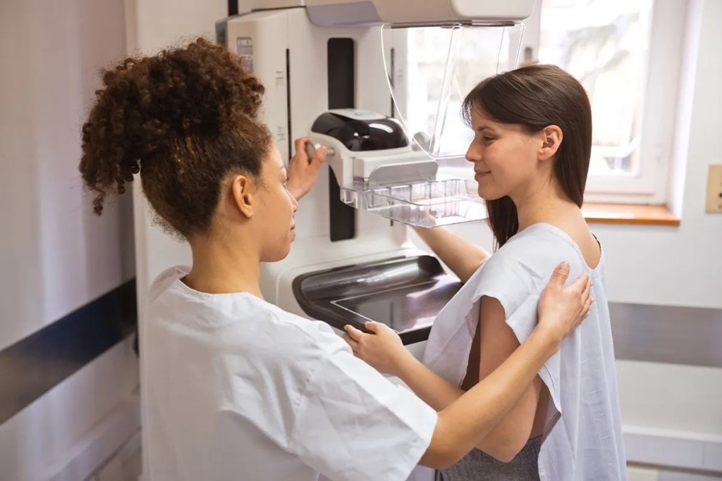
You will usually be asked to undress from the waist up and change into a gown before proceeding to the mammography machine. It is convenient to wear a top that is easy to remove, rather than a dress or jumpsuit, to make it easier to take your clothes off down to your waist. It is also important to remove any necklaces and nipple piercings.
If you’re nervous, tell the radiographer. If you’ve had a painful mammogram before, tell them. If you have a particular area that concerns you, point it out. That information is always useful.
During The Mammogram
Before your mammogram begins, there are a few things you can tell your technologist (the person operating the mammogram machine) to make the process and results smoother.
Communicate with your technologist:
- If you have breast implants. The technologist will take extra views of the breast (known as implant displacement views) if you have breast implants. This is to make sure all of your original breast tissue is viewable on the mammogram.
- If you’ve recently been vaccinated. Some people who receive a vaccine can develop swollen or enlarged lymph nodes that can show up on a mammogram. The radiologist will be able to more accurately interpret your mammogram result if they know that the swelling may be from a vaccine.
- If you have any tattoos on your arms, chest, or back. The ink could migrate to your lymph nodes and show up as white specks on a mammogram.
- If you need any accommodations – for example – if you have a disability that would make it difficult for you to stand or hold still during the mammogram, ask ahead of time about what accommodations they could provide.
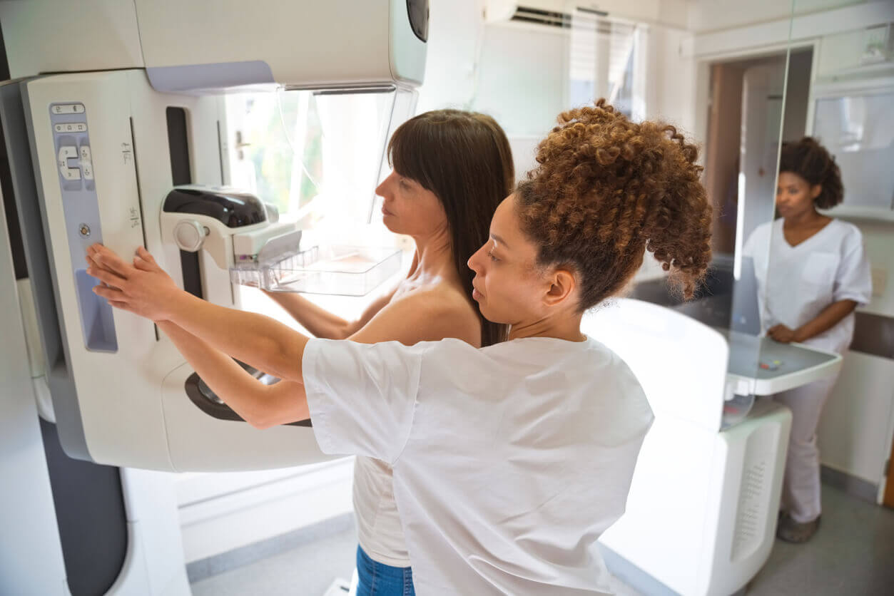
The mammogram itself is relatively quick. A radiographer will position you next to the mammography machine and place your breast onto the imaging platform. The breast is then gently but firmly compressed between two plates.
Compression is probably the part most women are curious — and sometimes anxious — about.
It can feel uncomfortable, particularly if your breasts are already tender. Some women describe it as pressure; others find it painful. If the compression becomes particularly painful to the point you cannot handle, tell the radiographer. They need enough compression to obtain a good-quality image, but you should not feel that you simply have to endure significant pain without saying anything.
The compression only lasts for a short period while each image is taken and it helps produce a clearer image while keeping the radiation exposure low. The reason for it is practical: compression spreads the breast tissue out, reduces movement and helps the X-ray image show small changes more clearly. It also allows the examination to use a relatively low radiation dose.
During the mammogram, the technologist will position each of your breasts in a mammography machine, one at a time. You will normally be asked to remain still and hold your breath briefly while each image is taken. Each breast will be compressed for only 20 – 30 seconds for each view.
Generally 2 views are obtained of each breast. The technologist may need to reposition you between images so that different areas of the breast tissue are captured.
If you have breast implants, the technique may be modified to obtain the best possible images of the breast tissue without damage to the implants or surrounding tissue.
The examination itself is fairly quick and is usually over within 10-20 minutes. The entire mammogram appointment takes about 30-40 minutes
What Happens Afterwards?
Once the images have been taken, a radiologist will review them. Depending on where you have the mammogram done, you may receive your report directly or it may be sent to the healthcare professional who referred you.
Sometimes you are told that everything looks reassuring and no further assessment is needed at that point. Sometimes the radiologist needs additional information or images.
You may be asked to return for:
- Additional mammogram views
- A breast ultrasound
- Comparison with previous mammograms
- Further assessment of a particular area
- Occasionally, a breast biopsy
Being asked to return does not automatically mean that cancer has been found.
There are many reasons why a screening mammogram may need a closer look. Sometimes tissue overlaps on the image. Sometimes a small area simply needs to be viewed from another angle. Sometimes the radiologist wants to compare it with an older mammogram to see whether it has changed.
This is one reason it is useful to have previous mammograms available when possible. Comparing current and previous images can help the radiologist determine whether an area is new or has remained stable.
What About The Results?
Your mammogram report may use a standard classification system called BI-RADS (Breast Imaging Reporting and Data System), which helps radiologists communicate what was seen and what follow-up, if any, is recommended.
You don’t need to memorise the categories. If your report contains unfamiliar terminology, ask your doctor or the radiology team to explain what it means in plain language. There is no shame in asking even the seemingly small questions about your health concerns.
A screening result is one part of the picture. Your symptoms, examination, previous imaging, personal risk and understanding all matter too.
What Does A Mammogram Actually Show?
A mammogram uses a small amount of X-ray radiation to create images of breast tissue. During the examination, your breast is positioned between two plates and compressed briefly while images are taken. These images are what determine the status of your exam.
Sometimes the mammogram is straightforward and you leave knowing that everything looks as expected. Sometimes the radiologist sees something that needs another look. That might mean another mammogram view, an ultrasound or, in some cases, a biopsy.

Breast cancer showing on a mammogram – Image courtesy of RSNA.
This is worth remembering before you go:
Being called back after a mammogram does not mean you have cancer.
Getting a mammogram is often considered the best way to find breast cancer early, when it’s most treatable. A mammogram can detect breast cancer before any symptoms develop. It can detect a tumor that cannot be felt. There are a variety of studies which have shown that having regular mammograms can lower your risk of dying from breast cancer.
A mammogram cannot be used to diagnose breast cancer on its own, though. Other tests are always needed before a diagnosis can be made. In most cases a biopsy is the only test that can tell for sure whether breast cancer is present, as well as the type.
Screening is designed to pick up changes that deserve a closer look. Most abnormalities found during screening do not turn out to be cancer, but they do need to be investigated properly. This is one of the reasons screening has both benefits and potential downsides, including false-positive results and additional testing. (Pace & Keating, 2014)
From The Maxima Consultation Room
There are questions women often ask once the door is closed and they can speak freely. Some are about mammograms. Others are about whether a particular breast change is “normal”.
These are some of the conversations we found were worth having.
“I’m only 40. Do I really need a mammogram already?”
This is a very reasonable question.
Age 40 isn’t a magic switch that suddenly makes breast cancer appear. Screening recommendations are based on how the likelihood of breast cancer changes with age and on the balance between finding cancers earlier and the possibility of false alarms and unnecessary investigations.
If you’re around 40, this is a good time to discuss your personal risk and screening options with your doctor rather than assuming that everyone needs exactly the same schedule.
If you have additional risk factors, your screening plan may be different.
“I’m not concerned because breast cancer doesn’t run in my family.”
We hear this one often. A family history of breast cancer can increase your risk, particularly when a close relative was diagnosed young or several relatives have had breast or ovarian cancer. Not having a family history doesn’t mean you’re not at risk.
Most women diagnosed with breast cancer do not have a known strong family history. Your family history is one piece of the picture. Your age, personal medical history and other risk factors matter too.
If you do have a strong family history, don’t wait until 40 to mention it. Your doctor may recommend a formal risk assessment or earlier and/or additional screening.
“If my mammogram is normal, that means I’m in the clear. right?”
A normal mammogram is reassuring. It isn’t a lifetime guarantee. Some breast cancers are difficult to see on mammography, and this can be particularly relevant in women with dense breast tissue.
Breast density describes the amount of fibrous and glandular tissue compared with fatty tissue in the breast. It has nothing to do with breast size. Dense breasts are common, especially among younger women, and they can make mammograms more difficult to interpret because dense tissue and some breast abnormalities both appear white on the X-ray.
That doesn’t mean everyone with dense breasts automatically needs additional scans. More imaging is not always better imaging. Additional tests can find things that are ultimately harmless but require further investigation. This is why breast screening is best treated as a conversation about risk, rather than a checklist of tests.
“My breasts are always lumpy. How will I know if something is actually different?”
Breast tissue can naturally feel lumpy or uneven, and hormonal changes can alter how your breasts feel throughout the menstrual cycle. The useful thing isn’t necessarily being able to identify every structure in your breast. It’s becoming familiar with what is normal for you.
A new lump, an area that feels distinctly different, a change that persists beyond your cycle, or another unusual change deserves assessment. You don’t need to decide whether it “feels like cancer”. That’s the healthcare professional’s job.
“I found a lump but my mammogram was normal. Should I still be worried?”
A normal mammogram is reassuring, but it doesn’t override a new physical finding. Some abnormalities, particularly in dense breast tissue, can be difficult to see on mammography.
If you can feel a persistent lump or have another concerning breast change, tell your doctor even if your recent mammogram was normal. Depending on your age and the clinical findings, further assessment such as an ultrasound or diagnostic mammogram may be appropriate.
Your symptoms still matter after a normal scan.
“Do I need an ultrasound as well as a mammogram?”
Not necessarily.
Ultrasound and mammography answer slightly different questions, and one does not simply replace the other in every situation. Ultrasound can be particularly useful when investigating a palpable lump or assessing certain findings on a mammogram. It can also provide additional information in women with dense breasts.
However, automatically adding every available scan to every mammogram isn’t necessarily better care. Additional imaging can lead to more false-positive findings, further investigations and sometimes biopsies. The decision should depend on your breast density, age, symptoms and overall risk.
“I have breast cancer in my family. Does that mean I need to start screening much earlier?”
Not necessarily — but it is something your doctor should know about.
The details of your family history matter. There is a difference between having one older relative who developed breast cancer and having several close relatives diagnosed at young ages, particularly if there is also a history of ovarian, pancreatic or prostate cancer or known genetic mutations in the family.
If your family history suggests an inherited risk, you may benefit from formal risk assessment and, in some circumstances, genetic counselling or testing. Don’t wait until your first routine mammogram to mention it. Bring it up when you are discussing your screening plan.
The Changes Worth Getting Checked
You don’t need to examine your breasts obsessively or follow a complicated monthly routine. What is useful is knowing what is normal for you.
Make an appointment if you notice a new or persistent:
- Lump or area of thickening
- Change in breast size or shape
- Dimpling, puckering or pulling of the skin
- Change in the nipple, including new inversion
- Unusual nipple discharge, especially bloody discharge
- Persistent redness or skin changes
- Swelling in the breast or armpit
- Change that doesn’t settle after your menstrual cycle
Most breast changes aren’t cancer. You shouldn’t have to work out which ones are harmless by yourself. That’s what your healthcare team is there for.
Early Detection Isn’t Just About Mammograms
There is sometimes a tendency to think of breast cancer screening as a single appointment every year or two. In reality, breast health is more layered than that.
It includes knowing your personal risk, being aware of changes in your breasts, having appropriate clinical examinations and using imaging when it is indicated.
South African data gives us another reason to take this seriously.
A 2025 study from two Johannesburg academic hospitals found that breast tumours diagnosed in 2022 were significantly smaller than those recorded at the same institutions in 2012. The researchers suggested that increased awareness, clinical breast examinations and improved detection may have contributed to this change, while also noting that the study represented a limited hospital population rather than South African women as a whole (De Lima et al., 2025)
The goal isn’t to find something because you’re expecting bad news. The goal is to make sure that if something is there, it has a chance of being found before it becomes harder to treat.
Your 40s are a good time to know your baseline. You don’t need to be frightened into looking after your breasts. You also don’t need to wait until you’re worried. If you’re approaching 40, ask your doctor about your personal breast cancer risk and what screening is appropriate for you. If you’re already over 40 and haven’t had that conversation, it’s not too late to have it.
If you’ve noticed a change in your breast — whether you’re 30, 40, 50 or 70 — don’t let your age or your last mammogram make the decision for you. A mammogram is a tool. Your breast health is the bigger picture.
The Maxima Takeaway
Knowing your breasts, understanding your risk and having the right screening at the right time can make breast health feel much less frightening — and much more manageable.
Ready to have the conversation?
Speak to us at Maxima Women’s Health or ask your healthcare provider about your personal breast cancer risk and whether mammography or another form of breast assessment is appropriate for you.
You don’t need to know what the change means. You just need to know when to ask about it.
Learn More About Your Breasts and Overall Health
Understanding what is normal for your body can make it easier to recognise when something needs attention. Read more about your health and well-being on our website.
Leave a Reply